PET Scan Appointments: Preparation, Imaging, and Results
Before an appointment for PET imaging, follow the imaging center’s instructions for food, activity, and medicines. Positron emission tomography uses a radioactive tracer and a special camera to show how tissues are functioning. PET imaging examinations usually include tracer administration, a waiting period, and time on the scanner table.
PET Scans and What They Show
PET stands for positron emission tomography. The tracer collects in tissues according to the biological process being studied, and the scanner detects signals produced as the radioactive material decays. A computer uses those signals to create images showing patterns of tracer uptake.
What is a PET scan used to assess? Depending on the tracer and clinical question, it can provide information about certain cancers, heart conditions, or brain disorders. For example, a commonly used tracer is related to glucose and helps show patterns of glucose use. The referring team chooses the examination for a particular question; uptake patterns require interpretation alongside other information.
Many PET scans are combined with computed tomography, or CT. PET provides functional information, while CT helps show where structures are located. A combined appointment may involve additional instructions or contrast material. The center can explain which parts of the examination are planned and why.
Preparing for the Appointment
Instructions vary with the tracer and examination. The center may ask for several hours without food and specify which drinks are allowed. It may also give instructions about exercise before the visit. Following the instructions matters because food, blood sugar, and activity can affect tracer distribution and image interpretation. If a preparation step was missed, call the center for advice before arriving.
Patients with diabetes should contact the ordering team and imaging center in advance for an individual plan for meals, blood sugar checks, and diabetes medicines. Do not change insulin or other prescribed medication without instructions from the treating team. Provide a complete list of medicines, vitamins, and supplements, including when they are normally taken.
Tell the team beforehand about possible or confirmed pregnancy, breastfeeding, recent illness, allergies, and relevant medical conditions. Ask about any special instructions for breastfeeding after the tracer and, if applicable, CT contrast. Mention difficulty lying still, pain, anxiety in enclosed spaces, or mobility needs so staff can plan support or discuss medication if appropriate.
Tracer and Waiting Period
At check-in, staff review preparation and may check blood glucose for an examination using a glucose-related tracer. They may ask the patient to change into a gown and remove metal objects that could affect images. An intravenous line is commonly placed in an arm or hand to give the radiopharmaceutical.
After injection, the tracer needs time to move through the body and collect in the tissues of interest. For many PET/CT examinations this takes about an hour, although timing depends on the tracer and purpose. The team may ask the patient to rest quietly during this period because muscle activity can affect some images. The center will explain whether water and a restroom visit are appropriate before scanning.
The tracer contains radioactive material, and the exposure depends on the examination. If CT is included, it adds radiation exposure. Questions about the expected exposure and the reason for the study belong with the ordering and imaging teams, who can explain the circumstances of the individual examination.
On the Scanner Table
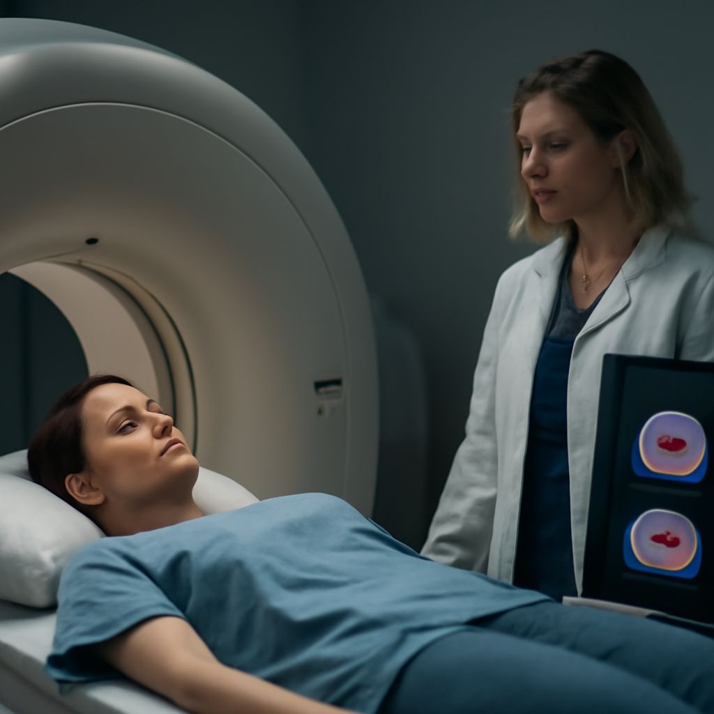
For the PET scan test, the patient generally lies on a narrow table that moves through a large ring-shaped scanner. The technologist positions the body and may use supports to make it easier to stay still. The camera detects signals from the tracer; it does not require the patient to feel or see them. Staff operate the scanner from another area and can communicate with the patient.
The patient may be asked to keep still and, for certain CT images, briefly hold a breath. The scan itself is generally painless, but remaining in one position can be uncomfortable. Tell staff about discomfort as soon as possible. The time on the table and total appointment length vary with the area, tracer, equipment, and whether additional images are needed; ask the center how long to allow.
After Imaging and Results
The team will provide instructions for eating, drinking, and resuming activities. It may recommend fluids and frequent urination to help clear the tracer, if appropriate for the patient. Follow any specific advice on contact with others or breastfeeding. If a sedative was given, ask about driving and arrange transportation as directed.
A physician trained in interpreting nuclear medicine images reviews the study and sends a report to the referring clinician. The report describes the findings in relation to the clinical question; an area of uptake can have more than one explanation. The ordering clinician considers the report alongside history, other tests, and previous imaging.
Before leaving, ask when the report should be available, who will discuss it, and whom to contact if it does not arrive. If further testing is mentioned, ask what question it would answer. RadiologyInfo describes the preparation, imaging process, and interpretation of PET/CT examinations.