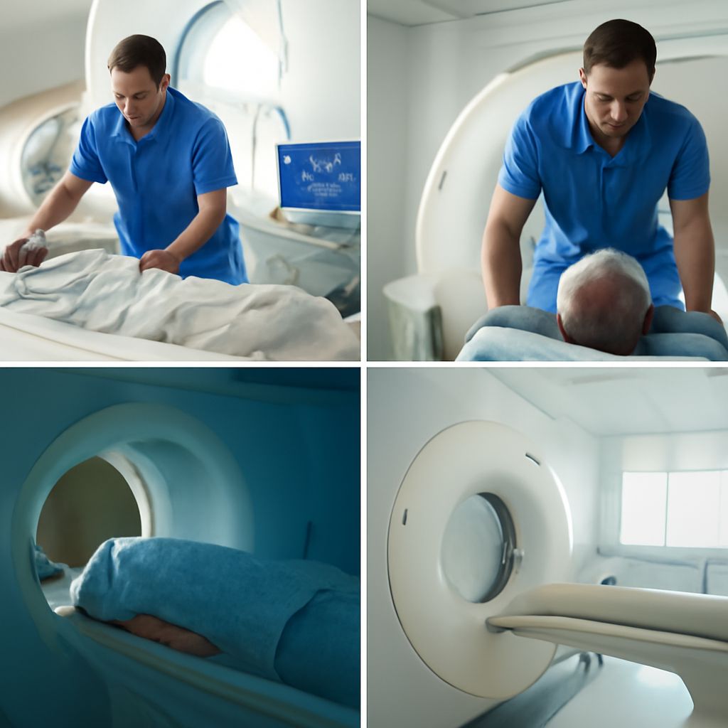
CT vs MRI: Understanding the Differences and Applications
In the realm of modern medicine, imaging tests have revolutionized the way practitioners diagnose and treat patients. From pinpointing soft tissue abnormalities to detecting bone fractures, these tests play a crucial role in patient care. Among the most frequently utilized are CT scans and MRIs. These imaging techniques offer distinct advantages, but understanding their differences is essential to determine the appropriate application for each. So, what can a CT scan show that an MRI cannot? This article delves into the nuances of each method, providing insights into specialized uses such as ovarian cancer MRI and the broader cancer MRI process.
Introduction to Imaging Tests
Medical imaging tests serve as the eyes of healthcare professionals, allowing them to see inside the body without invasive procedures. CT (Computed Tomography) scans and MRI (Magnetic Resonance Imaging) are among the most common tests, each providing unique insights. While both can create detailed images, their technological foundations differ, leading to varied uses.
CT scans use X-rays to generate images, making them particularly useful for examining bone injuries and detecting lung and chest problems. In contrast, MRIs employ magnetic fields and radio waves, excelling in visualizing soft tissues like the brain and muscles. Each method has its own pros and cons, making the choice between CT vs MRI situation-dependent.
CT vs MRI: Key Differences
The primary distinction between CT and MRI lies in their imaging techniques. CT scans excel in speed and are often used in emergency situations to quickly assess trauma and internal injuries. They are particularly adept at visualizing bone structures and detecting bleeding.
On the other hand, MRIs provide superior detail for soft tissues. They are invaluable for diagnosing conditions related to the brain, spinal cord, and muscles. The absence of radiation in the MRI process is another distinct advantage, especially for patients requiring frequent imaging.
While CT might be the go-to for assessing acute injuries, MRI is preferred for investigating complex joint disorders and neurological conditions. Thus, understanding when to utilize each can significantly impact patient outcomes.
What Can a CT Scan Show That an MRI Cannot?
Despite the versatility of MRIs, certain scenarios necessitate the use of CT scans. For instance, CT scans are highly effective in identifying bone fractures and calcifications that MRIs might miss. The ability to quickly capture images makes CT indispensable in emergency medicine, where time is of the essence.
CT scans also offer a better assessment of lung conditions. They can detect minor lung nodules and subtle changes in the pulmonary structure, which might not be as visible with MRI. Moreover, in detecting acute internal bleeding, CT’s rapid imaging is unmatched.
While MRI provides clearer images of soft tissues, CT scans are unparalleled in specific scenarios like evaluating complex bone structures. Hence, both imaging types have their own specialized applications.
Understanding Ovarian Cancer MRI
When it comes to diagnosing ovarian cancer, MRI plays a pivotal role. The ability of MRI to produce high-resolution images of soft tissues makes it particularly useful in assessing ovarian masses. An ovarian cancer MRI can help distinguish between benign and malignant tumors, guiding treatment decisions.
Unlike CT, MRI provides enhanced contrast resolution, allowing for better visualization of the ovaries and surrounding tissues. This detail is critical for staging cancer and planning surgeries or other interventions.
While CT scans may be used for initial assessments, MRIs offer comprehensive insights that are crucial for formulating a precise treatment plan for ovarian cancer patients.
Cancer MRI: A Comprehensive Look
Cancer diagnosis and treatment rely heavily on precise imaging. MRI is an invaluable tool in this regard, offering detailed visualization of tumors and surrounding tissues. The cancer MRI process can assess the size, extent, and sometimes even the type of cancer.
One of the significant benefits of MRI in cancer treatment is its ability to monitor tumor response to therapy, aiding in the adjustment of treatment plans. The technology’s superior soft tissue contrast ensures that even small changes in tumor size can be detected early.
Although CT scans remain a staple in oncology for staging and assessing metastatic spread, MRI provides complementary information that is often critical for a thorough understanding of the cancer’s progression and response to treatment.
Bone Scan vs MRI: When to Use Each
Both bone scans and MRIs provide crucial insights into musculoskeletal conditions, yet their applications differ significantly. Bone scans are nuclear imaging tests that highlight areas of increased bone activity, useful for detecting fractures, infections, and cancer spread.
In contrast, MRIs give detailed images of bones and surrounding soft tissues, making them ideal for diagnosing soft tissue injuries, disc herniations, and joint abnormalities. For precise localization and detailed assessment, MRI often surpasses bone scans, especially in musculoskeletal medicine.
Deciding between a bone scan and an MRI depends on the clinical scenario, with bone scans offering a broader view of bone activity and MRIs providing detailed anatomical images.
Bottom line: The choice between CT and MRI depends heavily on the clinical question at hand. Each imaging modality has unique strengths that cater to different diagnostic needs. By understanding these differences, healthcare providers can make informed decisions, ensuring accurate diagnosis and effective treatment planning.