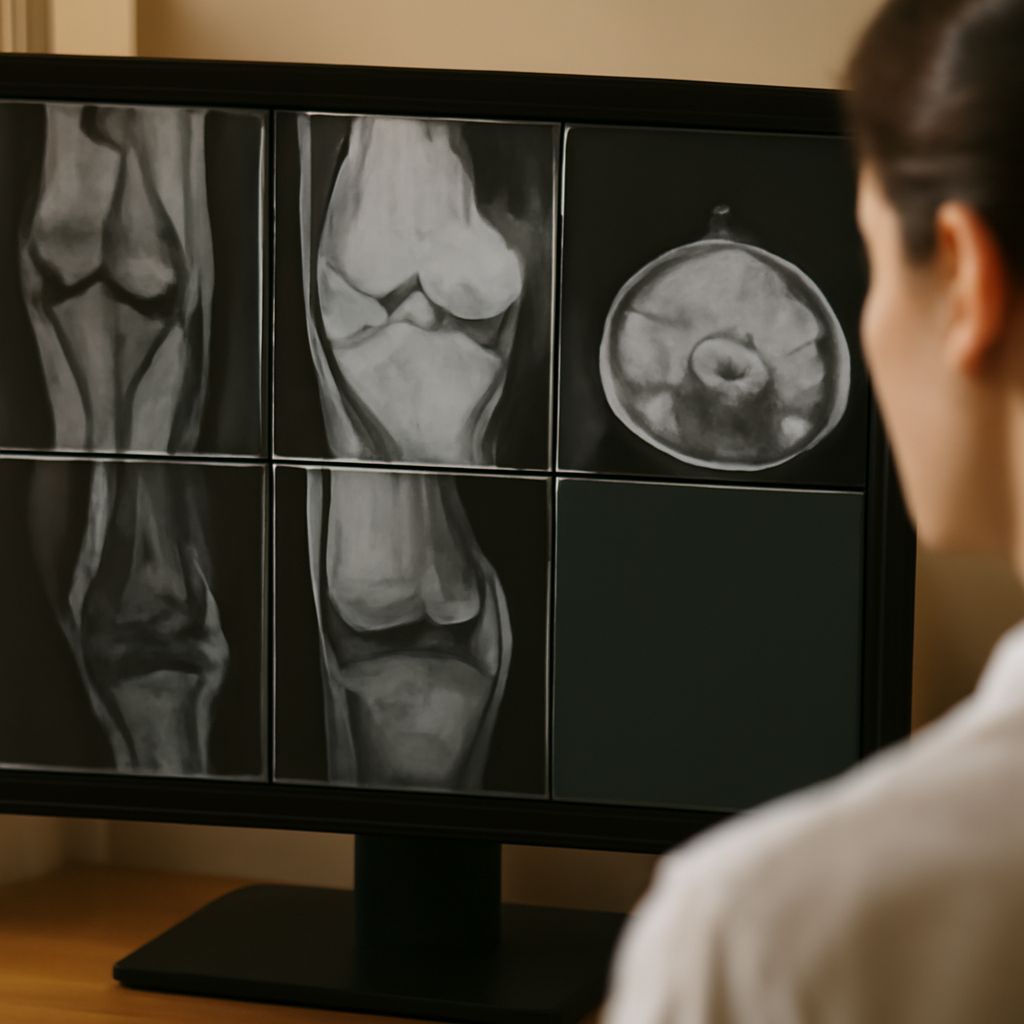
MRI vs CT Scan: Understanding the Differences
The realm of medical imaging is a nuanced landscape crucial for accurate diagnosis and treatment planning. Whether it’s deciphering a complex injury or monitoring the progression of a disease, selecting the right imaging test can significantly impact patient care. While MRI vs CT scan debates often arise, understanding the differences between these imaging modalities is vital. Additionally, knowing when to choose between an X-ray and an MRI can further refine diagnostic precision, ensuring optimal outcomes for patients.
Introduction to Medical Imaging
Medical imaging is an indispensable tool in modern healthcare, offering a window into the human body without the need for invasive procedures. Techniques like MRI (Magnetic Resonance Imaging), CT (Computed Tomography), and X-ray each have unique strengths, making them suitable for various diagnostic purposes. These imaging methods allow healthcare professionals to visualize internal structures, assess organ function, and identify abnormalities with remarkable clarity.
While each modality provides valuable insights, choosing the correct test depends on various factors, including the specific medical condition, the area of the body being examined, and the level of detail required. Understanding how these imaging techniques differ is essential for healthcare providers and patients alike.
MRI vs CT Scan: Key Differences
When considering an MRI versus a CT scan, several critical distinctions arise. MRI uses magnetic fields and radio waves to generate detailed images, making it ideal for soft tissue evaluation, such as brain, muscle, and joint assessments. In contrast, a CT scan employs X-rays to produce cross-sectional images, which are particularly useful for examining bony structures and detecting acute injuries.
The non-ionizing nature of MRI makes it a preferred choice for patients who require multiple scans, avoiding the cumulative radiation exposure associated with CT scans. However, MRI scans are typically lengthier and more expensive, factors that may influence the decision-making process. In emergency situations where rapid diagnosis is crucial, CT scans offer the advantage of speed, delivering results in a matter of minutes.
Ultimately, the choice between an MRI and a CT scan involves weighing the need for detailed imagery against the urgency and cost considerations. Each method serves its unique role, tailored to the specific diagnostic context.
Xray vs MRI: Comparative Insights
Comparing X-rays and MRIs reveals further distinctions in imaging capabilities. X-rays are a staple in medical imaging due to their efficiency and ability to quickly capture images of bones and certain chest conditions. However, they fall short in visualizing soft tissues, where MRIs excel.
MRIs provide unparalleled detail for soft tissue evaluation, making them the gold standard for neurological and musculoskeletal imaging. Patients with suspected ligament tears or brain anomalies might benefit more from an MRI than an X-ray, which cannot adequately capture these complexities.
Despite their limitations, X-rays are invaluable for initial assessments and follow-ups in specific cases, such as tracking bone healing. When deciding between an X-ray and an MRI, clinicians consider the specific diagnostic needs, balancing the thoroughness of MRI scans with the accessibility of X-rays.
Difference Between MRI and Xray
The primary difference between an MRI and an X-ray lies in their imaging approaches and applications. MRI is a non-invasive, non-radiative technique using magnetic fields to create detailed images of internal structures, optimal for soft tissues. This makes it ideal for conditions involving the brain, spine, joints, and blood vessels.
Conversely, X-rays work by passing radiation through the body to capture images, primarily of bones and other dense structures. They are less effective for soft tissues but excel in detecting fractures and infections in bones. The simplicity and speed of X-rays make them a go-to for initial diagnostic imaging, particularly in trauma cases.
Understanding the complementary roles of MRIs and X-rays helps in selecting the appropriate imaging modality based on clinical requirements, ensuring a balanced approach to patient care.
Choosing the Right Imaging Test
Determining the most suitable imaging test involves several considerations. Patient history, symptomatology, and the specific diagnostic question guide the choice between MRI, CT, or X-ray. MRI is often favored for its detailed soft tissue images, while CT scans are selected for their rapid execution and effectiveness in identifying acute conditions.
X-rays serve as the first line of investigation for bone assessments and certain thoracic issues, offering a quick and reliable diagnostic tool. In situations where detailed imaging of soft tissues is necessary, MRI becomes the preferred option despite its higher cost and longer acquisition time.
Healthcare providers must weigh the benefits and limitations of each modality, integrating imaging results with clinical findings to formulate a comprehensive diagnostic strategy that best supports patient outcomes.
Safety Recap: When employing imaging tools like CT scans and MRIs, consider the balance between radiation exposure and diagnostic benefit. MRI’s non-radiative nature offers a safer alternative for repeat imaging, while CT scans provide rapid, high-detail results essential for urgent diagnoses.