Pelvic Ultrasound: What to Expect, How to Prepare, and What It Shows
If your doctor ordered a pelvic ultrasound, knowing what the scan involves and how your body position affects image quality will help you walk in prepared. Pelvic ultrasound prep requirements vary depending on whether the scan is transabdominal or transvaginal, and skipping the preparation step leads to poor-quality images that often require a repeat visit. For women, an ovaries ultrasound is one of the most common reasons this imaging is ordered, though the pelvis contains several structures the scan evaluates simultaneously. Understanding how thyroid ultrasound images explained compare to pelvic imaging can help patients who need both procedures understand what each one actually shows. Whether your provider specified transabdominal or transvaginal, good pelvic ultrasound preparation takes about 30–60 minutes of planning before you arrive.
This article breaks down what pelvic ultrasound shows, how to prepare correctly, and what to expect during the exam itself.
What a Pelvic Ultrasound Evaluates
A pelvic ultrasound uses sound waves to produce real-time images of the organs in the lower abdomen and pelvis. In women, the scan evaluates the uterus, cervix, fallopian tubes, and ovaries. In men, it examines the bladder, prostate gland, and seminal vesicles. Providers order pelvic imaging to investigate pelvic pain, abnormal bleeding, urinary symptoms, and suspected growths or cysts.
Sonographers look for structural abnormalities such as uterine fibroids, endometrial polyps, ovarian cysts, and signs of ectopic pregnancy. For urinary concerns, the bladder wall and post-void residual volume are measured. Evaluating the prostate via pelvic sonography can detect enlargement or suspicious areas that warrant further workup. The scan takes 15–30 minutes and produces no radiation exposure.
Ovaries Ultrasound: What the Imaging Shows
When the primary focus is the ovaries, the sonographer measures each ovary and documents the size, shape, and any follicles or cysts present. An ovarian ultrasound identifies simple cysts, complex masses, polycystic ovary morphology, and free fluid around the ovaries that could indicate rupture or torsion. Doppler flow assessment is often added to evaluate blood supply to ovarian masses, which helps differentiate benign from potentially concerning lesions.
Ovarian imaging looks different than thyroid imaging despite both relying on ultrasound technology. While thyroid ultrasound images show nodule echogenicity, calcifications, and vascularity in a superficial gland, ovarian imaging involves deeper structures where image quality depends heavily on bladder fullness and body habitus. The principles of ultrasound interpretation are the same, but the anatomy and clinical questions are entirely different.
Pelvic Ultrasound Prep: How to Prepare Before Your Scan
For a transabdominal pelvic ultrasound, you typically need a full bladder. The standard instruction is to drink 32 ounces of water about one hour before the exam and avoid urinating until after the scan. A full bladder pushes the bowel out of the pelvic field and creates a fluid window that improves visualization of pelvic structures. Arriving with an inadequate bladder fill is the most common reason for suboptimal transabdominal pelvic studies.
For a transvaginal pelvic ultrasound, you will be asked to empty your bladder before the exam, not fill it. The transvaginal probe is placed inside the vaginal canal and sits adjacent to the structures being imaged, so bladder distension is not necessary and can actually worsen visualization. Preparing for a pelvic ultrasound transvaginally means arriving with a comfortably empty bladder and understanding that a chaperone will be present.
Some providers order both types in sequence. In that case, you start with a full bladder for the abdominal portion, then empty your bladder for the transvaginal portion. Confirm which type your order specifies before you begin drinking water, since misunderstanding the preparation can delay your appointment.
Types of Pelvic Ultrasound and What to Expect During the Exam
Transabdominal pelvic sonography involves lying on an exam table while a technologist applies gel to your lower abdomen and moves a handheld transducer across the skin surface. Pressure may be applied to improve visualization. The exam is painless, though a full bladder creates significant discomfort that resolves immediately after you void.
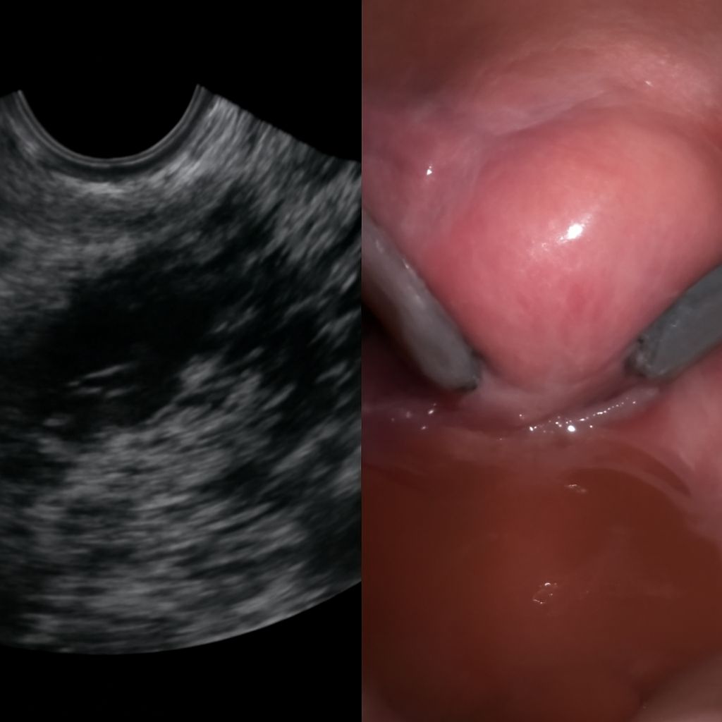
Transvaginal ultrasound involves a thin, covered probe inserted a few centimeters into the vaginal canal. Patients insert the probe themselves or the sonographer does so with your guidance. The procedure feels similar to a gynecological speculum exam and is typically better tolerated than patients expect. Images taken transvaginally show significantly more detail than transabdominal images for most pelvic structures because the probe sits closer to the target anatomy.
Key takeaways: follow the prep instructions specific to the type of scan ordered, arrive on time so your bladder fill is accurate, and ask your provider which structures will be evaluated so you can anticipate what the results will address.