Thyroid Histology and Gland Tissue: A Practical Overview
Histology, the microscopic study of tissue structure, gives clinicians and students a window into how organs function at the cellular level. Thyroid histology reveals the characteristic follicular architecture that makes the gland instantly recognizable under the microscope. Alongside it, thymus histology labeled diagrams and thyroid gland histology labeled slides form the foundation of endocrine histology coursework in medical and allied health programs.
Understanding uterus histology labeled and prostate gland histology alongside thyroid tissue broadens the comparative picture of glandular organs. Each has a distinct architecture tied directly to its secretory function. This overview focuses on thyroid structure while placing it in context with other commonly studied endocrine and reproductive glands.
Thyroid Histology: Follicles and Colloid
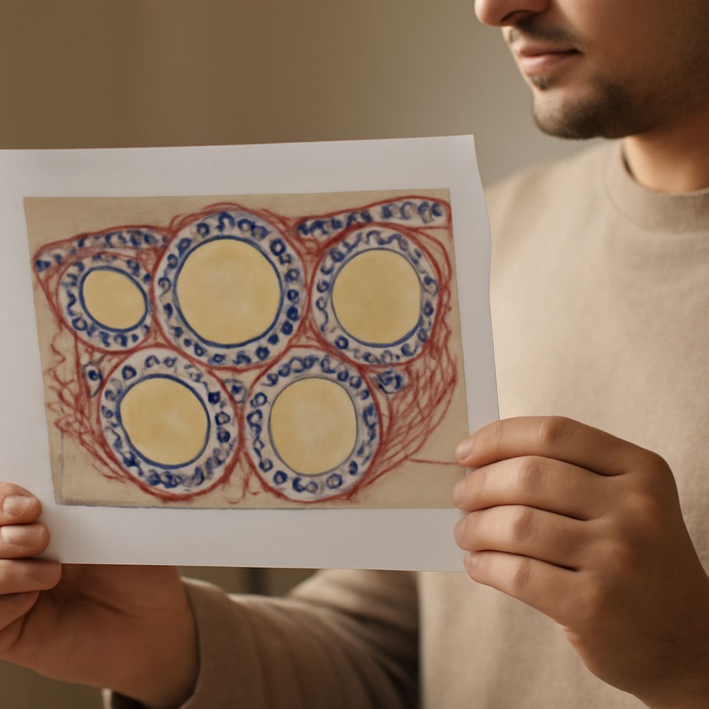
The defining feature of thyroid histology is the follicle. Each follicle is a spherical structure lined by a single layer of follicular epithelial cells, called thyrocytes, surrounding a lumen filled with colloid. Colloid is a glycoprotein-rich material composed primarily of thyroglobulin, the precursor to thyroid hormones T3 and T4.
In normally functioning thyroid tissue, follicles vary in size and the epithelium is cuboidal. When the gland is hyperactive, cells become columnar and colloid diminishes as hormones are released. In hypothyroid states, follicles enlarge and colloid accumulates. These histological changes correspond directly to the functional state of the gland.
Parafollicular C cells scattered between follicles produce calcitonin, a hormone involved in calcium regulation. They are not visible in routine hematoxylin and eosin staining without special techniques but are an important component of thyroid gland histology labeled for complete anatomical understanding.
Thymus Histology Labeled: Structure and Immune Function
Thymus histology labeled diagrams typically show the outer cortex and inner medulla of each thymic lobule. The cortex is densely packed with immature T lymphocytes undergoing selection. The medulla contains Hassall’s corpuscles, concentric whorls of epithelial cells unique to the thymus and used as a histological landmark.
The thymus is most active in childhood and undergoes involution after puberty, with lymphoid tissue replaced progressively by fat. In histology courses, thymus histology labeled slides appear alongside thyroid gland histology labeled images to contrast lymphoid with endocrine architecture.
Uterus Histology Labeled: Layers and Cycle Changes
Uterus histology labeled diagrams identify three main layers: the endometrium, myometrium, and perimetrium. The endometrium is the innermost glandular layer that undergoes cyclic changes driven by estrogen and progesterone. It is divided into the functional layer, which is shed during menstruation, and the basal layer, which regenerates it.
The myometrium is thick smooth muscle providing the contractile force for labor. Uterine histology changes dramatically across the menstrual cycle, making it a useful model for studying hormone-driven tissue remodeling in comparison to the relatively stable architecture of thyroid histology.
Prostate Gland Histology: Glands and Stroma
Prostate gland histology shows compound tubuloalveolar glands embedded in a fibromuscular stroma. The epithelium is typically pseudostratified columnar. Corpora amylacea, laminated concretions found in the glandular lumens, are a characteristic feature that increases in frequency with age.
Prostate histology is clinically significant because adenocarcinoma of the prostate is graded using the Gleason system, which relies entirely on glandular architecture under the microscope. Comparing normal prostate gland histology with pathological changes gives pathologists diagnostic criteria for one of the most common male malignancies.