How to Read an MRI: A Patient-Friendly Guide to MRI Images
Waiting for MRI results can be stressful, and many patients want to understand what the images show before their follow-up appointment. Knowing how to read an MRI at a basic level does not replace professional radiology interpretation, but it does help you engage more meaningfully with your healthcare team and understand the language used in your report. A working knowledge of MRI image reading principles transforms a confusing set of gray-scale images into a comprehensible picture of your anatomy.
This guide explains how to read MRI images, covers the fundamentals of how to read a MRI report, provides tips for reading an MRI with confidence, and also addresses the practical question of cost for MRI scan across different settings.
The Basics: How to Read an MRI Image
MRI images appear as gray-scale pictures where different tissues reflect magnetic resonance signals at different intensities. The fundamental skill in how to read an MRI at any level is understanding what appears bright (hyperintense) versus dark (hypointense) and why. Unlike X-rays—which primarily show density—MRI shows tissue characteristics based on water content, fat content, and molecular environment.
Tissue Contrast on T1 and T2 Sequences
MRI generates multiple image sequences, each optimized to highlight different tissue characteristics. On T1-weighted images, fat appears bright (white), water appears dark (gray-black), and most soft tissues appear in intermediate shades of gray. T1 images are excellent for evaluating anatomy and fat-containing structures. On T2-weighted images, water appears bright and fat appears gray or dark. T2 sequences highlight fluid-containing structures—edema, cysts, and cerebrospinal fluid all appear prominently bright. Most MRI studies include both T1 and T2 sequences to provide complementary information about normal and abnormal tissues.
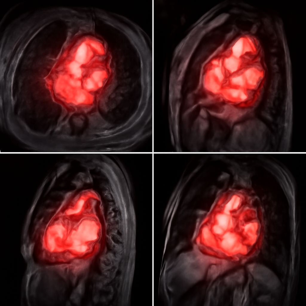
Planes of View: Axial, Coronal, Sagittal
MRI can generate images in three standard planes. Axial images are cross-sectional slices viewed from below (as if you were looking up through the body). Coronal images show front-to-back slices as seen from the front. Sagittal images provide side-view slices as if viewed from the side. Radiologists examine all three planes when reading a study, as lesions may be more clearly visible or better characterized in one orientation than another. When reviewing your own MRI images, confirming which plane you are looking at helps you orient to the anatomy correctly.
Reading an MRI: Common Findings Explained
When reading an MRI scan, certain findings appear consistently across different body regions. A dark, round, well-defined area on T2 within solid tissue often represents a cyst (fluid-filled structure), which is almost always benign. Bright signal on T2 within or around bone typically indicates edema or inflammation. Enhancement after gadolinium contrast injection—where a structure that was dark before contrast becomes bright after it—indicates that the blood-brain barrier (in brain MRI) or vascular permeability is disrupted, which can signify tumors, abscesses, or active inflammation.
Disc herniations on spine MRIs appear as protrusions of darker disc material pressing against the brighter cerebrospinal fluid surrounding the spinal cord or nerve roots. Meniscal tears in knee MRIs show as linear bright signal within the normally dark meniscal tissue on certain sequences. These pattern-recognition principles are the foundation of radiological interpretation—the same logic of brightness and signal intensity applies across body regions and pathology types.
Understanding Your MRI Report as a Patient
MRI reports are written by radiologists for referring physicians, using precise anatomical and clinical terminology. When reviewing your report, focus on the Impression or Conclusion section, which summarizes the key findings in plain language. The Findings section provides detailed descriptions of each structure examined and may include technical terminology that requires clarification.
Common terms to know: unremarkable means normal; no acute findings means nothing requiring immediate attention; nonspecific means the finding does not point to a definitive diagnosis; correlate clinically means the radiologist is asking the ordering physician to interpret the imaging finding in the context of the patient’s symptoms. If any term in your MRI report is unclear, asking your ordering physician or a nurse navigator to explain it is entirely appropriate—and encouraged.
Cost for MRI Scan: What to Expect and How to Save
The cost for MRI scan varies significantly by facility type, body region, geographic location, and whether contrast is used. Hospital outpatient departments typically charge $1,000 to $3,000 or more for a single MRI. Independent outpatient imaging centers—which often use the same equipment operated by equally qualified technologists—frequently charge $400 to $1,200 for equivalent studies. The price gap between hospital-based and independent facility MRI pricing is one of the most dramatic examples of healthcare cost variation in the U.S. system.
Patients with insurance should confirm their deductible status and whether the imaging center is in-network before scheduling. Self-pay patients should ask directly for the cash-pay rate, which many centers offer at a discount. Comparing MRI prices across local providers through your insurer’s cost transparency tool or independent healthcare cost comparison sites can identify significant savings without compromising diagnostic quality. Always ensure the facility you choose is accredited by the American College of Radiology (ACR) or an equivalent body to confirm quality standards.
Key takeaways: Understanding MRI images begins with learning how T1 and T2 signal characteristics distinguish tissue types, and how the three imaging planes provide complementary views of anatomy. MRI report terminology can be decoded by focusing on the Impression section and asking your provider to explain unfamiliar terms. Comparing imaging facilities for MRI pricing can produce substantial savings without sacrificing diagnostic quality.