Cardiac Ultrasound Views: A Comprehensive Guide
Cardiac Ultrasound Views: A Comprehensive Guide
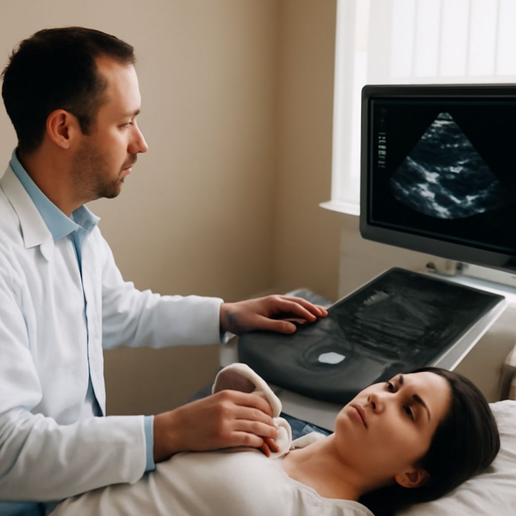
The field of cardiology has seen remarkable advances, with cardiac ultrasound views being pivotal in diagnosing and monitoring heart conditions. A parasternal long axis ultrasound is indispensable in cardiac assessments, providing detailed images that aid in the evaluation of heart structures. Moreover, understanding axis deviation EKG is vital for interpreting electrical activity and diagnosing potential cardiac issues. Ultrasound biomicroscopy offers insights into cardiac anatomy at a microscopic level, enhancing diagnostic precision.
Understanding Cardiac Ultrasound Views
Introduction to Cardiac Ultrasound
Cardiac ultrasound, commonly known as echocardiography, utilizes sound waves to create images of the heart. This non-invasive technique offers a dynamic view of heart functioning, essential for diagnosing various cardiac conditions.
Importance of Cardiac Ultrasound Views
The different views obtained during cardiac ultrasound provide comprehensive insights into heart anatomy and function. These perspectives are integral for identifying structural anomalies and assessing myocardial performance.
Common Cardiac Ultrasound Techniques
Several techniques are employed in cardiac ultrasound, including transthoracic echocardiography and transesophageal echocardiography. These methods differ in approach but both yield crucial information about heart health.
Parasternal Long Axis Ultrasound
Overview of Parasternal Long Axis
The parasternal long axis view is a staple in echocardiographic evaluation. This perspective allows for detailed visualization of the left ventricle and surrounding structures, essential for comprehensive cardiac assessment.
Techniques for Accurate Imaging
Achieving precise imaging in the parasternal long axis requires skillful positioning of the transducer and careful interpretation of the images. This technique is critical for measuring cardiac dimensions and assessing valve function.
Clinical Applications
Parasternal long axis ultrasound is frequently used to evaluate cardiac function and detect abnormalities such as hypertrophy or valvular disease. It is a fundamental part of routine echocardiographic exams.
Axis Deviation EKG Insights
Understanding Axis Deviation
Axis deviation refers to the orientation of the heart’s electrical activity. This concept is vital for interpreting EKG results and can indicate underlying cardiac conditions, such as conduction abnormalities.
EKG Analysis Techniques
EKG analysis involves assessing the electrical signals of the heart, with axis deviation being a key factor. This analysis helps uncover arrhythmias and other cardiac disorders, providing a roadmap for treatment.
Clinical Significance
The clinical significance of axis deviation lies in its ability to reveal potential cardiac issues. Detecting deviations early can lead to timely interventions, improving patient outcomes significantly.
Ultrasound Biomicroscopy in Cardiology
What is Ultrasound Biomicroscopy?
Ultrasound biomicroscopy employs high-frequency sound waves to produce detailed images of cardiac tissues at a microscopic level. This advanced imaging technique allows for thorough examination of heart morphology.
Applications in Cardiology
In cardiology, ultrasound biomicroscopy is used to investigate cardiac structures with exceptional precision. This approach is particularly beneficial for assessing small, intricate anatomical details.
Advanced Imaging Techniques
Advanced imaging methods such as ultrasound biomicroscopy enhance diagnostic capabilities by providing clear, detailed views of cardiac tissues, thereby aiding in accurate diagnosis and treatment planning.
AFI Ultrasound and Its Role
AFI Ultrasound Basics
AFI ultrasound, or Amniotic Fluid Index ultrasound, although primarily used in obstetrics, has implications in cardiology through advanced imaging techniques that can adapt for cardiac evaluation.
Clinical Applications
In clinical settings, AFI ultrasound demonstrates potential in cardiac assessments by providing detailed imaging that can be adapted from its traditional use in prenatal care to cardiology.
Comparisons with Other Ultrasound Methods
When compared to other ultrasound methods, AFI ultrasound offers a unique perspective with its adaptable imaging techniques, making it a versatile tool in the cardiologist’s toolkit.
Bottom line: Cardiac ultrasound views, including parasternal long axis ultrasound, axis deviation EKG, and ultrasound biomicroscopy, are indispensable in modern cardiology. They provide vital insights that enhance diagnosis and intervention strategies, ensuring better patient care.