Abnormal Thyroid Ultrasound: Results, Normal Comparisons, and Next Steps
Abnormal Thyroid Ultrasound: Results, Normal Comparisons, and Next Steps
Receiving a report that flags irregularities in your thyroid imaging can feel alarming, but most abnormal thyroid ultrasound findings require monitoring rather than immediate intervention. An ultrasound of the thyroid gland is the primary imaging tool for evaluating nodules, cysts, and structural changes, and interpreting results requires comparing what the scan shows against established normal parameters. A normal thyroid ultrasound shows a butterfly-shaped gland with uniform echotexture, clearly defined borders, and no nodules or masses. When your report deviates from that baseline, understanding what the thyroid ultrasound results mean is the first step toward an informed conversation with your physician.
Many patients also wonder what does ovarian cancer look like on an ultrasound, reflecting a broader concern about how ultrasound imaging detects malignancy in different organs. While the thyroid and ovary are distinct structures with different imaging criteria, both benefit from standardized assessment frameworks that guide clinical decisions. This article focuses on thyroid-specific findings and how normal vs abnormal thyroid ultrasound criteria apply to real-world reports.
What Normal Thyroid Ultrasound Findings Include
Size, Echogenicity, and Texture
A normal thyroid ultrasound typically shows a gland measuring roughly 4 to 5 cm in length per lobe, with a smooth capsule and homogeneous internal echogenicity. The parenchyma should appear slightly brighter than the surrounding strap muscles. Vascularity on Doppler imaging is present but not exaggerated. Small colloid cysts — smooth, anechoic pockets — are common incidental findings that do not indicate pathology.
When Findings Fall Within Acceptable Limits
Mild asymmetry between thyroid lobes and tiny sub-centimeter nodules with purely cystic characteristics often fall within normal limits for clinical management purposes. The American Thyroid Association risk stratification system classifies nodules by their ultrasound features, and many low-risk nodules are simply followed over time without biopsy. Understanding thyroid scan results requires knowing these classification frameworks, which most radiology reports reference.
Interpreting Abnormal Thyroid Ultrasound Features
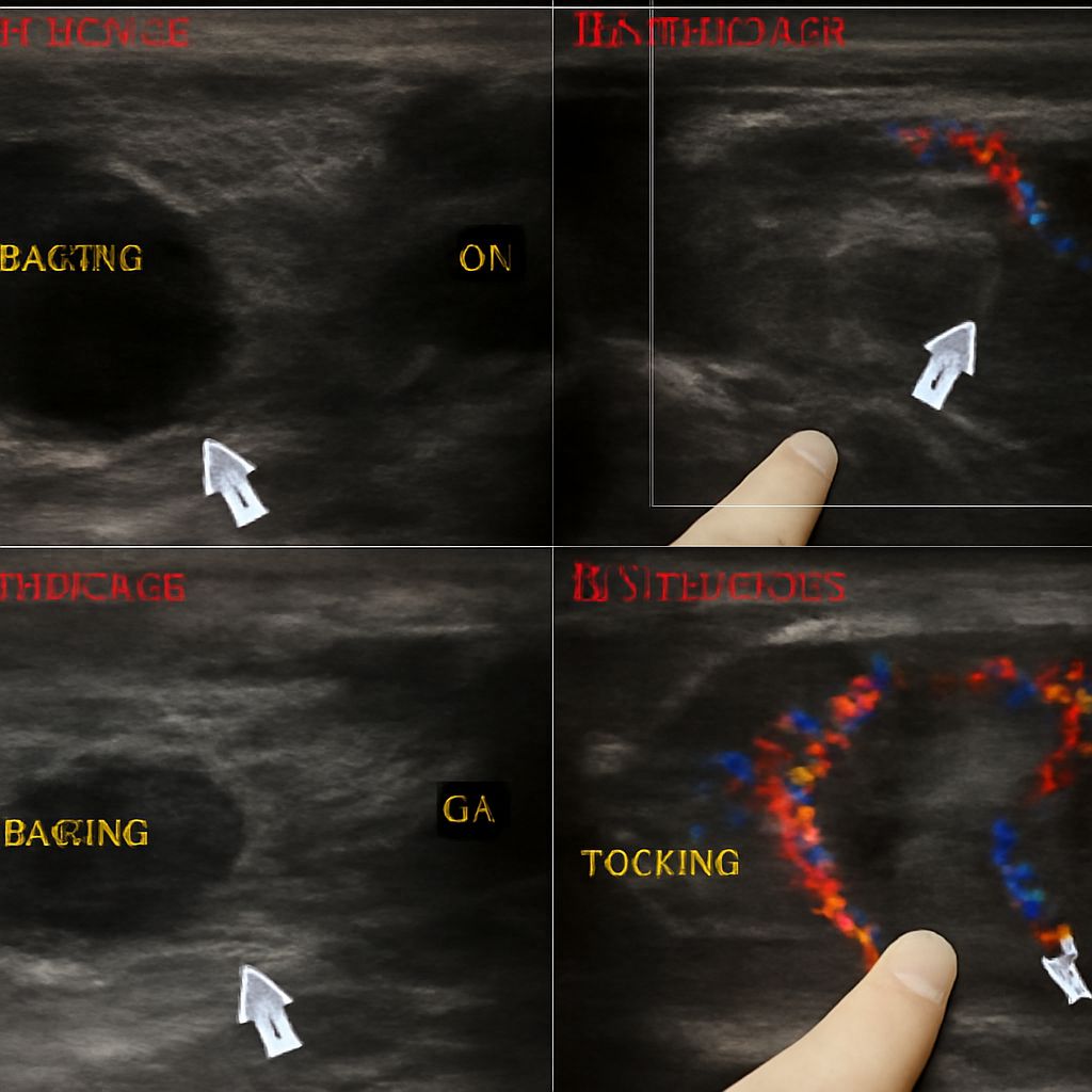
Findings that suggest a higher-risk nodule include hypoechogenicity relative to surrounding thyroid tissue, irregular or microlobulated margins, taller-than-wide shape, microcalcifications (tiny bright spots), and increased internal vascularity on Doppler. Thyroid ultrasound results that check several of these boxes typically prompt a fine-needle aspiration biopsy to obtain cells for pathology review. A report noting these features is not a diagnosis of cancer — it is a prompt for further evaluation using established clinical criteria.
Comparing normal vs abnormal thyroid ultrasound features side by side helps patients understand why certain nodules are biopsied and others are not. A well-differentiated cystic nodule with smooth walls carries a very different risk profile than a solid, hypoechoic mass with calcifications.
Ovarian Cancer on Ultrasound vs. Thyroid Findings
Patients curious about what does ovarian cancer look like on an ultrasound will find that ovarian malignancy has its own distinct imaging features: complex cystic masses with thick septations, solid components, papillary projections, or internal vascularity. These criteria differ entirely from thyroid nodule assessment, though the principle of systematic risk stratification applies to both. In both cases, ultrasound findings guide — but do not replace — biopsy and pathology in confirming or excluding malignancy.
After an Abnormal Thyroid Report: What Happens Next
Your endocrinologist or primary care provider will review your thyroid imaging results in the context of your symptoms, thyroid function tests (TSH, free T4), and any prior imaging. High-risk nodule features typically lead to a biopsy referral within weeks. Intermediate-risk findings may prompt a follow-up ultrasound in 6 to 12 months to check for growth or feature changes. Low-risk findings often require no action beyond routine monitoring.
Next steps: Request a copy of your full radiology report and bring it to your endocrinology appointment. Ask your provider which risk tier your nodule falls into, what the recommended follow-up interval is, and under what circumstances a biopsy would be added to the plan.