Six Week Ultrasound: What to Expect and Beyond
Six Week Ultrasound: What to Expect and Beyond
The journey of pregnancy is filled with anticipation, with each week unveiling new wonders and challenges. Among the earliest milestones, the six week ultrasound plays a crucial role. If you’re curious about what to expect, including insights into the 6 week fetus ultrasound and the nuances of urgent care X-rays, this guide is for you. Understanding these early imaging tests can provide reassurance and clarity during this pivotal time.
While the ultrasound at 7 weeks offers more detailed views of early fetal development, the six week ultrasound sets the foundation. It’s essential to recognize when urgent care X-rays might be necessary, ensuring both maternal and fetal health. Let’s explore the specifics and expectations surrounding these early stages.
Understanding the Six Week Ultrasound
What is a Six Week Ultrasound?
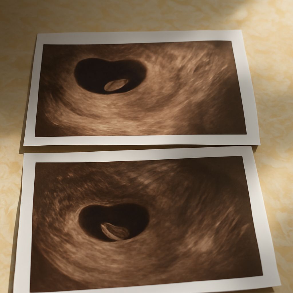
A six week ultrasound is an early pregnancy scan typically conducted to confirm the pregnancy and check basic embryonic development. It involves using sound waves to create a visual representation of the developing embryo, allowing healthcare providers to assess early signs of fetal growth.
During this scan, the technician will look for the presence of a gestational sac and fetal pole, and in some cases, even an early heartbeat. This crucial step helps in determining the viability of the pregnancy.
Benefits of a Six Week Ultrasound
The advantages of a six week ultrasound include early detection of potential issues and confirmation of the estimated due date. By analyzing the early stages of fetal development, healthcare providers can identify any abnormalities early on.
Moreover, the scan provides peace of mind for expectant parents by confirming the presence of the embryo. This early glimpse into the womb can alleviate anxieties and set a positive tone for the rest of the pregnancy journey.
Comparing Ultrasound at Different Stages
Ultrasound at 4 Weeks
At four weeks, an ultrasound might not reveal much detail. Typically, only the gestational sac might be visible, as the embryo is minuscule and still in its formative stages. This scan is usually conducted to confirm the presence of an intrauterine pregnancy.
Due to the limited information at this stage, healthcare providers often recommend waiting until the six week ultrasound for more comprehensive insights.
Ultrasound at 7 Weeks
By seven weeks, ultrasonography provides a more detailed view. The embryo is more developed, often allowing for the visualization of a heartbeat. This stage can offer a clearer picture of the pregnancy’s progression and viability.
The ultrasound at 7 weeks is particularly valuable for verifying fetal health and ensuring the pregnancy is progressing as expected. It builds upon the findings of the six week ultrasound, offering a richer understanding of the embryo’s development.
What to Expect During a 6 Week Fetus Ultrasound
Preparing for the Ultrasound
Preparation for a six week fetus ultrasound is straightforward. Patients are typically advised to drink water before the procedure to fill the bladder, enhancing image quality. A full bladder helps in achieving better visualization during the scan.
Comfort is key, so wearing loose-fitting clothes can ease the process. Being mentally prepared for the possibility of limited results is also important, as this is an early stage of pregnancy.
Interpreting the Results
Results from a six week ultrasound can be varied. If a heartbeat is detected, it is a positive sign of a viable pregnancy. However, the absence of a heartbeat at this early stage does not necessarily indicate a problem, as it might simply be too early.
Healthcare providers will interpret the findings and provide guidance on next steps, which may include further ultrasounds to monitor progress. Understanding the nuances of this early scan can help manage expectations and guide future medical decisions.
When to Consider Urgent Care X-Rays
Situations Requiring Urgent Care X-Rays
While ultrasounds are the primary imaging tool during early pregnancy, there are situations where urgent care X-rays may be necessary. This includes cases of trauma or suspected injury, where an X-ray can provide crucial information without focusing on the fetus.
It is important to consult with healthcare providers to weigh the risks and benefits, as radiation exposure is generally minimized during pregnancy.
Differences Between Ultrasounds and X-Rays
Ultrasounds and X-rays serve different purposes. While ultrasounds use sound waves to visualize soft tissues, X-rays utilize radiation to image bones and denser structures. Ultrasounds are preferred for fetal imaging due to their safety profile.
X-rays might be considered in specific medical scenarios and are typically conducted with protective measures to safeguard the fetus. Understanding these differences ensures informed decisions during pregnancy.
Pro tips recap: Early pregnancy scans, such as the six week ultrasound, provide essential insights into fetal development. Always consult healthcare providers for personalized advice. Prepare adequately for appointments, and understand the potential need for urgent care X-rays in certain situations. This knowledge empowers you to navigate early pregnancy with confidence.