Mole Biopsy: What to Expect and How to Care for the Site
Mole Biopsy: Procedure Types, Healing, and What Results Mean
A mole biopsy is performed when a skin lesion has features that raise concern for dysplasia or melanoma — asymmetry, irregular borders, color variation, large diameter, or evolution over time. Punch biopsy scar formation is common and usually minor, but proper aftercare minimizes visible scarring. Skin cancer biopsy results determine whether additional surgery is needed, how much margin must be cleared, and what the ongoing monitoring plan should be. Skin biopsy aftercare for a mole removal site involves daily wound care until healing is complete. Redness around skin biopsy site is expected initially, but expanding redness after the first 48 hours warrants a clinical evaluation.
Understanding what happens during and after a mole biopsy helps patients follow aftercare instructions and interpret results accurately.
Types of Mole Biopsy Techniques
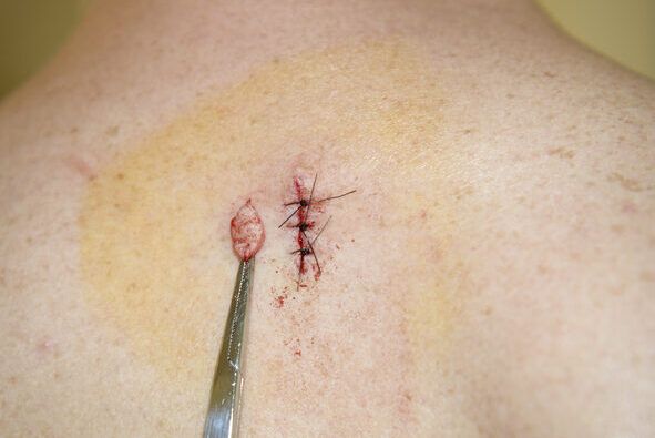
Shave biopsy removes a thin slice of the lesion without full-thickness skin removal — appropriate for raised lesions where depth extension is unlikely. Punch biopsy uses a cylindrical instrument to remove a core of skin including deeper dermal tissue — used when full-thickness histology is needed to assess invasion depth. Excisional biopsy removes the entire lesion with a margin of normal skin — used for lesions strongly suspicious for melanoma where complete removal is both diagnostic and potentially therapeutic. The technique chosen affects the resulting punch biopsy scar size, shape, and healing timeline.
Skin Biopsy Aftercare for Mole Sites
After a mole biopsy, the site should be kept clean and moist. Clean the wound gently once daily with mild soap and water. Apply a thin layer of petrolatum-based ointment and cover with a non-adherent dressing. Avoid soaking the wound in pools, baths, or oceans until fully healed. Sun protection over the healing site prevents hyperpigmentation. Punch biopsy sutures should not be submerged and must stay clean and dry between care sessions. Most punch biopsy sites with sutures heal within two weeks; shave biopsy sites within one to two weeks depending on size and location.
Interpreting Skin Cancer Biopsy Results
Pathology reports from a skin cancer biopsy describe the tissue type, the presence of dysplasia or malignancy, and the margins. A report showing melanoma in situ means cancer cells are confined to the epidermis. Invasive melanoma reports include Breslow depth, which measures how deeply cancer cells have penetrated — this directly affects surgical margins required and whether sentinel lymph node biopsy is recommended. Benign results — common blue nevi, dysplastic nevi with mild atypia — may require periodic monitoring rather than further surgery.
Key takeaways: Mole biopsy results dictate next steps — clarify with your dermatologist what your specific pathology report means for surveillance and any additional procedures. Keep your aftercare consistent and sun-protect the site to minimize scar formation and pigment changes.